

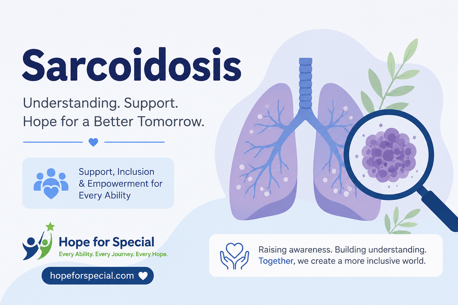
Sarcoidosis: The Hidden Disease That Mimics Everything & What You Can Do About It in 2026 🎗️
A sarcoidosis diagnosis often comes after months — sometimes years — of frustrating dead ends. One day it is “probably asthma.” The next it is “possibly lupus.” Then a biopsy finally reveals the truth. If that journey sounds familiar, you are not alone. Sarcoidosis is one of the most underdiagnosed and misunderstood diseases in modern medicine. 💙
This guide gives you everything. The biology, the symptoms doctors miss, the 2026 treatment options, real patient stories, and the expert insights that most websites never share.

- ⚡ What Is Sarcoidosis? — Direct Answer
- 🔬 Understanding Sarcoidosis More Deeply
- 🧬 What Causes Sarcoidosis?
- 🫁 Types of Sarcoidosis & Organs Affected
- Pulmonary Sarcoidosis 🫁 (Most Common — ~90% of cases)
- Cutaneous Sarcoidosis 🩹 (~25% of cases)
- Ocular Sarcoidosis 👁️ (~25% of cases)
- Cardiac Sarcoidosis ❤️ (~5–10% of diagnosed cases; possibly up to 25% subclinically)
- Neurosarcoidosis 🧠 (~5–10% of cases)
- Hepatic & Splenic Sarcoidosis 🫀
- Other Organs Affected
- 🩸 Symptoms of Sarcoidosis — Early & Advanced
- 🔬 How Is Sarcoidosis Diagnosed?
- Step 1 — Clinical History & Physical Examination
- Step 2 — Blood Tests
- Step 3 — Imaging
- Step 4 — Tissue Biopsy (Usually Required)
- Step 5 — Pulmonary Function Tests (PFTs)
- 📊 Stages of Pulmonary Sarcoidosis
- 💊 Sarcoidosis Treatment Options in 2026
- When Is Treatment NOT Required?
- First-Line Treatment — Corticosteroids
- Second-Line Treatments (Steroid-Sparing Agents)
- Third-Line Treatment — Biologic Agents
- Treatment for Specific Organ Involvement
- 📈 Key Sarcoidosis Statistics
- 🌿 Prognosis & What to Realistically Expect
- 💛 A Patient’s Real Story: “Ten Years. Fourteen Doctors. One Answer.”
- 🔍 What You Must Not Skip About Sarcoidosis — Expert Insights
- 1. 😴 Fatigue in Sarcoidosis Is Not “Just Tiredness” — It Is a Disease in Itself
- 2. 🧬 Small Fiber Neuropathy — The Painful Secret of Sarcoidosis
- 3. 🏃 Exercise Is Medicine — Not Something to Avoid
- 4. 🌞 Vitamin D and Calcium — A Misunderstood Relationship
- 5. 🧠 Cognitive Symptoms Are Real and Common
- 6. ❤️ Cardiac Screening Is Critically Underpracticed
- ⏳ Milestones in Sarcoidosis Research
- ❓ FAQs
- Can sarcoidosis go away on its own without treatment?
- Is sarcoidosis an autoimmune disease?
- What does sarcoidosis fatigue feel like?
- Can sarcoidosis cause death?
- How long does sarcoidosis last?
- Can sarcoidosis affect pregnancy?
- Is sarcoidosis related to lupus?
- What should I eat if I have sarcoidosis?
- What is the difference between sarcoidosis and tuberculosis?
- Can sarcoidosis affect children?
- 🔗 Trusted Resources for Sarcoidosis Patients & Families
- ✨ Final Thoughts: You Are Not Alone in the Sarcoidosis Journey
⚡ What Is Sarcoidosis? — Direct Answer
Sarcoidosis is a chronic inflammatory disease where clusters of immune cells form small lumps called granulomas in organs throughout the body. It most commonly affects the lungs and lymph nodes. It can also involve the skin, eyes, heart, and brain. Many patients recover without treatment. Others need long-term management. Early diagnosis saves lives.
🔬 Understanding Sarcoidosis More Deeply
To truly understand sarcoidosis, you need to understand what a granuloma is — because granulomas are the defining feature of this disease.
Normally, when the immune system detects a foreign substance it cannot destroy — a bacteria, fungus, or unknown irritant — it surrounds it with immune cells to wall it off. This cluster of cells is called a granuloma. In most cases, once the threat is gone, the granuloma dissolves.
In sarcoidosis, this process goes wrong. The immune system forms granulomas even when there is no clearly identified threat — and these granulomas do not dissolve. Instead, they persist, accumulate, and over time can disrupt the normal function of whatever organ they form in.
The lungs are involved in approximately 90% of sarcoidosis cases. But sarcoidosis is genuinely a whole-body disease. It can form granulomas in virtually any organ — and this is precisely why it is so difficult to diagnose and so easy to misidentify as something else entirely.
💡 Voice Search Answer — What is sarcoidosis in simple terms? Sarcoidosis is a disease where the immune system overreacts and forms small clusters of inflamed cells called granulomas in the body’s organs. The lungs are most commonly affected. In many cases, sarcoidosis gets better on its own. In others, it becomes chronic and requires treatment to prevent organ damage.
🧬 What Causes Sarcoidosis?
This is the question that frustrates both patients and physicians most. The honest answer in 2026 remains: we do not know for certain. But we know far more than we did a decade ago.
The Leading Theories
1. Immune System Dysregulation
The most widely accepted theory holds that sarcoidosis develops when a genetically susceptible person’s immune system encounters a specific trigger — likely an environmental antigen — and mounts an exaggerated, self-sustaining inflammatory response. The immune system essentially loses the ability to turn itself off.
2. Environmental & Occupational Triggers
Research has consistently linked sarcoidosis to certain environmental exposures:
- 🌿 Organic dust (mold, hay, bird droppings)
- 🏭 Inorganic particles (silica, beryllium, aluminum)
- 🌳 Mycobacteria and propionibacteria (bacteria found in soil and skin)
- 🏥 Healthcare and first-responder work environments
The landmark World Trade Center Health Study found a significantly elevated rate of sarcoidosis among rescue and recovery workers exposed to Ground Zero dust — providing strong evidence that environmental triggers can initiate the disease in susceptible individuals.
3. Genetic Predisposition
Sarcoidosis is not directly inherited, but genetic susceptibility plays a clear role. Certain HLA (Human Leukocyte Antigen) gene variants — particularly HLA-DRB1 — are significantly overrepresented in sarcoidosis patients. First-degree relatives of sarcoidosis patients have a 3–5 times higher risk of developing the disease compared to the general population.
4. Infectious Agents
Multiple studies have detected mycobacterial and propionibacterial antigens within sarcoidosis granulomas. This has led some researchers to propose that sarcoidosis may represent an abnormal immune response to specific microbial triggers rather than a true autoimmune disease. This distinction has important treatment implications.
Who Is Most at Risk?
⚠️ Age group: Most commonly diagnosed between ages 25–40 and again after 50
⚠️ Race: African Americans are 3–4 times more likely to develop sarcoidosis than white Americans — and tend to have more severe disease
⚠️ Gender: Women are slightly more commonly affected than men, particularly for cardiac and neurological sarcoidosis
⚠️ Occupation: Healthcare workers, firefighters, agricultural workers, and metal workers have elevated rates
⚠️ Geography: Higher rates in Scandinavia, Northern Europe, and among African Americans in the Southern United States
✅ Not contagious — Sarcoidosis cannot be spread from person to person
🫁 Types of Sarcoidosis & Organs Affected
Sarcoidosis can affect virtually any organ. Understanding which organs are involved drives every treatment decision.
Pulmonary Sarcoidosis 🫁 (Most Common — ~90% of cases)
The lungs are the most frequent target. Granulomas form in lung tissue and around bronchi. Over time, they can cause scarring (pulmonary fibrosis) that permanently reduces lung function. Symptoms include:
- Persistent dry cough
- Shortness of breath
- Chest tightness or discomfort
- Wheezing (less common)
Cutaneous Sarcoidosis 🩹 (~25% of cases)
The skin is the second most commonly affected organ. Presentations include:
- Erythema nodosum — Painful red nodules on the shins. Actually a sign of acute, self-limiting sarcoidosis with good prognosis.
- Lupus pernio — Violaceous (purple-red) plaques on the nose, cheeks, and ears. A marker of chronic, systemic sarcoidosis.
- Maculopapular rash — Small raised skin lesions, often around scars or tattoos.
Ocular Sarcoidosis 👁️ (~25% of cases)
Eye involvement is common and can be sight-threatening if untreated. The most frequent manifestation is uveitis — inflammation of the middle layer of the eye. Symptoms include eye redness, pain, light sensitivity, and blurred vision. Regular ophthalmology monitoring is essential for all sarcoidosis patients.
Cardiac Sarcoidosis ❤️ (~5–10% of diagnosed cases; possibly up to 25% subclinically)
Cardiac sarcoidosis is one of the most dangerous and underdiagnosed forms. Granulomas in the heart’s electrical conduction system can cause:
- Life-threatening arrhythmias
- Complete heart block
- Sudden cardiac death
- Heart failure
⚠️ Critical Point: Many cases of cardiac sarcoidosis are discovered only at autopsy in patients who died of sudden cardiac death and were never diagnosed with sarcoidosis during their lifetime. This is why cardiac screening (MRI and PET scan) is now recommended for all newly diagnosed sarcoidosis patients.
Neurosarcoidosis 🧠 (~5–10% of cases)
When granulomas form in the nervous system, the results can include:
- Facial nerve palsy (drooping face)
- Headaches and meningitis-like symptoms
- Cognitive changes and memory problems
- Seizures
- Peripheral neuropathy (numbness, tingling in hands and feet)
- Diabetes insipidus (from pituitary involvement)
Hepatic & Splenic Sarcoidosis 🫀
The liver and spleen are involved in approximately 50–80% of sarcoidosis cases — but most cases are subclinical (no symptoms). Symptomatic liver involvement is less common but can cause elevated liver enzymes, abdominal discomfort, and rarely liver failure.
Other Organs Affected
| Organ | Approximate Frequency | Key Manifestation |
|---|---|---|
| Lungs | ~90% | Cough, breathlessness, fibrosis |
| Lymph nodes | ~90% | Enlarged nodes (especially chest) |
| Skin | ~25% | Rashes, nodules, lupus pernio |
| Eyes | ~25% | Uveitis, dry eyes, vision loss |
| Liver | ~50–80% (subclinical) | Elevated enzymes, rarely failure |
| Heart | ~5–25% | Arrhythmias, heart block |
| Nervous system | ~5–10% | Facial palsy, seizures |
| Kidneys | ~5% | Elevated calcium, kidney stones |
| Bones & joints | ~5–10% | Arthritis, bone lesions |
| Parotid glands | ~5% | Facial swelling |
🩸 Symptoms of Sarcoidosis — Early & Advanced
Sarcoidosis earns its nickname as “the great imitator” because its symptoms overlap with so many other conditions. This is why the average time from first symptom to diagnosis is 3–5 years — one of the longest diagnostic delays of any chronic disease.
Early Symptoms (Often Misattributed)
- 😴 Profound fatigue — The most common and debilitating symptom. Often dismissed as stress or depression.
- 🌡️ Low-grade fever — Persistent mild fever without obvious infection
- ⚖️ Unexplained weight loss
- 😮💨 Shortness of breath — Especially with exertion
- 😣 Chest discomfort or tightness
- 👁️ Red, painful, or blurry eyes
- 🩹 Skin rashes — Particularly on the shins (erythema nodosum)
- 🦵 Joint pain and swelling — Often in the ankles
Advanced Symptoms
- 🫁 Severe breathing difficulty from pulmonary fibrosis
- ❤️ Palpitations, fainting, or chest pain from cardiac involvement
- 🧠 Neurological symptoms: facial drooping, memory problems, seizures
- 🦷 Dry mouth and eyes (from salivary gland involvement)
- 💧 Excessive thirst and urination (from hypercalcemia or diabetes insipidus)
- 😰 Severe, treatment-resistant fatigue affecting all daily activities
Löfgren Syndrome — A Special Presentation
Löfgren syndrome is an acute form of sarcoidosis with a distinctive clinical picture:
✅ Erythema nodosum (painful skin nodules on shins)
✅ Enlarged lymph nodes in the chest (bilateral hilar lymphadenopathy)
✅ Fever and joint pain
✅ Excellent prognosis — approximately 90% of Löfgren syndrome cases resolve on their own within 2 years without specific treatment
Recognizing Löfgren syndrome is important because it often does not require aggressive treatment and carries a much better outcome than other forms of sarcoidosis.
🔬 How Is Sarcoidosis Diagnosed?
Diagnosing sarcoidosis requires a combination of clinical findings, imaging, and tissue biopsy. There is no single blood test that confirms sarcoidosis. This is part of why it is so frequently and frustratingly delayed.
Step 1 — Clinical History & Physical Examination
A thorough history of symptoms, occupational exposures, family history, and organ system involvement provides the first framework for suspicion. Physical examination may reveal enlarged lymph nodes, skin lesions, eye changes, or abnormal breath sounds.
Step 2 — Blood Tests
No single blood test diagnoses sarcoidosis, but several support the evaluation:
| Test | What It Indicates in Sarcoidosis |
|---|---|
| Serum ACE (Angiotensin-Converting Enzyme) | Elevated in ~60% of active sarcoidosis cases; useful for monitoring activity |
| Serum calcium | Elevated in ~10–15% of cases; from granuloma vitamin D production |
| 24-hour urine calcium | More sensitive than serum calcium for detecting hypercalciuria |
| Liver function tests | Elevated in hepatic sarcoidosis |
| Complete blood count | May show lymphopenia (low lymphocytes), anemia |
| Inflammatory markers (ESR, CRP) | Elevated in active disease |
| Lysozyme | Elevated in some cases; less specific than ACE |
Step 3 — Imaging
- Chest X-ray — First-line. Classically shows bilateral hilar lymphadenopathy (enlarged lymph nodes on both sides of the chest). Chest X-ray findings stage pulmonary sarcoidosis (Stages 0–IV).
- High-resolution CT (HRCT) scan — Provides far greater detail. Shows characteristic patterns including perilymphatic nodules, ground glass opacities, and fibrotic changes.
- PET scan — Identifies metabolically active granulomas throughout the body. Essential for cardiac sarcoidosis evaluation and guiding biopsy sites.
- Cardiac MRI — Gold standard for detecting cardiac sarcoidosis when arrhythmia or conduction abnormalities are present.
Step 4 — Tissue Biopsy (Usually Required)
Tissue biopsy demonstrating non-caseating granulomas (granulomas without central necrosis) is the cornerstone of sarcoidosis diagnosis — after other causes of granulomatous disease (particularly tuberculosis and fungal infections) are excluded.
Biopsy sites chosen are the safest and most accessible positive sites:
- Skin lesions — If present, safest biopsy site
- Peripheral lymph nodes — Easy access, good yield
- Endobronchial biopsy via bronchoscopy — Very high yield (~70–90%) for pulmonary sarcoidosis
- EBUS (Endobronchial Ultrasound) — Minimally invasive; allows biopsy of chest lymph nodes with ~90% diagnostic yield
- Liver, heart, or brain biopsy — Reserved for cases where safer sites are negative
Step 5 — Pulmonary Function Tests (PFTs)
Spirometry and diffusing capacity tests quantify the degree of lung function impairment. They are essential for monitoring disease progression and treatment response in pulmonary sarcoidosis. A restrictive pattern with reduced DLCO (diffusing capacity) is the classic finding.
📊 Stages of Pulmonary Sarcoidosis
Pulmonary sarcoidosis is classified into five radiological stages based on chest imaging findings:
| Stage | Chest X-Ray Findings | Spontaneous Remission Rate |
|---|---|---|
| Stage 0 | Normal chest X-ray (extrapulmonary disease only) | N/A |
| Stage I | Bilateral hilar lymphadenopathy only | ~60–80% |
| Stage II | Bilateral hilar lymphadenopathy + lung infiltrates | ~50–60% |
| Stage III | Lung infiltrates without hilar lymphadenopathy | ~30% |
| Stage IV | Pulmonary fibrosis (irreversible scarring) | ~0% (irreversible) |
Source: American Thoracic Society — Sarcoidosis Statement
💡 Important: These stages are radiological descriptions — they do not perfectly predict symptoms or quality of life. Many Stage II patients feel well. Some Stage I patients have severe fatigue and extrapulmonary disease. Staging must always be combined with clinical assessment.
💊 Sarcoidosis Treatment Options in 2026
Treatment of sarcoidosis is not one-size-fits-all.

The decision to treat depends on which organs are involved, how severely they are affected, and whether the disease is progressing.
When Is Treatment NOT Required?
Remarkably, many sarcoidosis patients do not need treatment. Spontaneous remission — where the disease resolves on its own without medication — occurs in:
- ~60–80% of Stage I pulmonary sarcoidosis patients
- ~50–60% of Stage II patients
- ~30% of Stage III patients
For patients with mild symptoms, normal or near-normal organ function, and no involvement of high-risk organs (heart, brain, eyes), a strategy of watchful waiting with regular monitoring is appropriate and recommended.
First-Line Treatment — Corticosteroids
Oral prednisone (or prednisolone) remains the mainstay of sarcoidosis treatment when treatment is needed. Corticosteroids suppress the immune response driving granuloma formation.
- Starting dose: typically 20–40mg prednisone daily
- Initial treatment duration: 3–6 months, then slow taper
- Maintenance may be needed for 12–24 months or longer in chronic disease
- Effective for most organ manifestations but carries significant long-term side effects with prolonged use
Second-Line Treatments (Steroid-Sparing Agents)
When corticosteroids cause intolerable side effects or fail to control the disease, steroid-sparing alternatives are used:
| Drug | Mechanism | Best Used For | Key Consideration |
|---|---|---|---|
| Methotrexate | Antimetabolite; immunosuppressive | Pulmonary, skin, joint sarcoidosis | Monitor liver function; requires folic acid supplementation |
| Azathioprine | Purine synthesis inhibitor | Pulmonary, neurological sarcoidosis | Check TPMT enzyme level before starting |
| Leflunomide | Pyrimidine synthesis inhibitor | Alternative to methotrexate | Teratogenic; strict contraception needed |
| Hydroxychloroquine | Antimalarial; anti-inflammatory | Skin, calcium-related, fatigue | Safe in pregnancy; less effective for lung disease |
| Mycophenolate | Lymphocyte proliferation inhibitor | Pulmonary, cardiac, neurological | Well tolerated; rising use in 2026 |
Third-Line Treatment — Biologic Agents
For patients with severe, refractory sarcoidosis who fail corticosteroids and steroid-sparing agents, biologic therapies targeting specific immune pathways offer meaningful benefit:
🔬 Infliximab (Remicade) — An anti-TNF monoclonal antibody. The best-studied biologic for sarcoidosis. Demonstrated significant improvement in FVC (forced vital capacity) and quality of life in controlled trials for pulmonary and cutaneous sarcoidosis.
🔬 Adalimumab (Humira) — Another anti-TNF agent. Used when infliximab is not tolerated. Evidence base is smaller but growing.
🧪 JAK inhibitors (tofacitinib, ruxolitinib) — An exciting emerging class in 2026. Early trials and case series show promise particularly for cutaneous and pulmonary sarcoidosis refractory to other treatments. Larger controlled trials are underway.
🧪 Rituximab — Anti-CD20 biologic targeting B-cells. Used in select cases of refractory neurosarcoidosis.
Treatment for Specific Organ Involvement
Cardiac Sarcoidosis:
- Corticosteroids to suppress active granulomatous inflammation
- Implantable Cardioverter-Defibrillator (ICD) for patients at risk of sudden death
- Permanent pacemaker for complete heart block
- Cardiac transplantation in end-stage cardiac sarcoidosis
Ocular Sarcoidosis:
- Topical corticosteroid eye drops for anterior uveitis
- Systemic corticosteroids or immunosuppressants for posterior uveitis
- Regular ophthalmology monitoring every 6–12 months
Neurosarcoidosis:
- High-dose corticosteroids (often IV methylprednisolone initially)
- Early addition of steroid-sparing agents (methotrexate, mycophenolate)
- Infliximab for refractory neurosarcoidosis
Managing Hypercalcemia:
- Sun avoidance and dietary calcium moderation
- Adequate hydration to protect kidney function
📈 Key Sarcoidosis Statistics
| Statistic | Figure | Source |
|---|---|---|
| Estimated sarcoidosis cases in the US | ~200,000 active cases | Foundation for Sarcoidosis Research |
| Annual new sarcoidosis diagnoses in the US | ~35,000–50,000 | American Thoracic Society |
| Global sarcoidosis prevalence (estimated) | 1–40 per 100,000 population | World Health Organization |
| Percentage of cases involving the lungs | ~90% | National Heart, Lung & Blood Institute |
| Spontaneous remission rate (all stages) | ~50–70% | European Respiratory Society |
| Average time from first symptom to diagnosis | 3–5 years | Foundation for Sarcoidosis Research |
| African American incidence vs. white Americans | 3–4x higher | NCI SEER Database |
| Percentage developing chronic sarcoidosis | ~30% | American Thoracic Society |
| Mortality rate from sarcoidosis (US) | ~1–5% | National Heart, Lung & Blood Institute |
| Cardiac sarcoidosis proportion of sarcoidosis deaths | ~13–25% | Heart Rhythm Society |
🌿 Prognosis & What to Realistically Expect
The prognosis of sarcoidosis is genuinely varied. This is not a disease with a single trajectory. Understanding the range of outcomes helps patients and families plan meaningfully.
Favorable Prognostic Signs
✅ Löfgren syndrome presentation
✅ Stage I or Stage II disease at diagnosis
✅ Erythema nodosum as presenting feature
✅ Acute onset with clear symptoms (rather than insidious discovery)
✅ Absence of cardiac, neurological, or Stage IV pulmonary involvement
✅ African American patients with acute presentation (paradoxically, despite higher severity rates overall)
Less Favorable Prognostic Signs
⚠️ Stage III or IV pulmonary disease at diagnosis
⚠️ Cardiac or neurological involvement
⚠️ Lupus pernio skin lesions (marker of chronic systemic disease)
⚠️ Progressive disease despite 6 months of treatment
⚠️ Multiple organ involvement at diagnosis
⚠️ Age over 40 at diagnosis
The Three Natural Courses of Sarcoidosis
1. Acute / Self-limiting (most common): Disease resolves on its own within 1–3 years. No long-term organ damage.
2. Chronic / Stable: Disease persists but remains stable without significant progression. Requires ongoing monitoring and sometimes long-term low-dose treatment.
3. Chronic / Progressive: Disease progresses despite treatment, leading to significant organ damage — particularly pulmonary fibrosis, heart failure, or neurological disability. This is the most challenging trajectory and the focus of the most active 2026 research efforts.
🎯 Key Context: Even for patients with chronic progressive sarcoidosis, new treatments — particularly biologic agents and JAK inhibitors — are changing outcomes. Patients who were previously treatment-refractory are achieving meaningful disease control with 2024–2026 therapeutic advances.
💛 A Patient’s Real Story: “Ten Years. Fourteen Doctors. One Answer.”
“My name is Diana. I was 32 when the fatigue started. Not tired — truly exhausted. As if someone had turned gravity up to twice its strength. Then came the cough. Then a skin rash on my shin that my GP called ‘probably eczema.’ Then my eyes went red and painful for weeks.
I saw a pulmonologist, a rheumatologist, a dermatologist, a gastroenterologist, and two neurologists over four years. I was told it was anxiety. I was told it was fibromyalgia. I was told it might be lupus. I was put on medications that did nothing or made things worse.
I cried in the car. Not because it was good news. But because finally — finally — someone knew what was wrong.
I was started on prednisone, then transitioned to methotrexate. My fatigue improved within four months. My breathing improved. My eye inflammation resolved. I have Stage II disease that has been stable for three years now.
What I wish I had known from the beginning: sarcoidosis is a real disease. The fatigue is real. The symptoms are real. Push for a specialist. Push for a chest CT. Push for a biopsy. Do not let anyone tell you it is anxiety until every box has been checked.”
— Diana M., sarcoidosis patient diagnosed 2019, stable since 2022 ✨
🔍 What You Must Not Skip About Sarcoidosis — Expert Insights
1. 😴 Fatigue in Sarcoidosis Is Not “Just Tiredness” — It Is a Disease in Itself
The fatigue experienced in sarcoidosis is unlike normal tiredness. It does not improve with rest. It is not proportional to activity. It is medically classified as cancer-related fatigue equivalent in severity in many sarcoidosis patients. Yet it is consistently undertreated because it is invisible on imaging and normal on routine blood tests.
New research in 2024–2026 has identified elevated serum TNF-alpha, reduced mitochondrial function, and autonomic nervous system dysregulation as contributing mechanisms. Patients should specifically raise fatigue with their sarcoidosis specialist — it is treatable with targeted strategies including low-dose naltrexone (LDN), structured exercise rehabilitation, and in some cases infliximab.
2. 🧬 Small Fiber Neuropathy — The Painful Secret of Sarcoidosis
A growing body of research confirms that small fiber neuropathy (SFN) is present in approximately 40% of sarcoidosis patients — yet most patients are never tested for it. SFN causes burning, tingling, and pain in the hands and feet that is frequently misattributed to anxiety or fibromyalgia.
Diagnosis requires a specialized skin punch biopsy measuring nerve fiber density — a test most general neurologists do not routinely perform. Identifying SFN in sarcoidosis changes the treatment approach significantly.
3. 🏃 Exercise Is Medicine — Not Something to Avoid
Many sarcoidosis patients are told by well-meaning clinicians to “take it easy” and “avoid exertion.” Current evidence, however, strongly supports the opposite. Pulmonary rehabilitation and structured supervised exercise programs consistently improve exercise capacity, fatigue levels, and quality of life in sarcoidosis patients — including those with significant pulmonary involvement.
Fear of exercise in sarcoidosis is common but usually unfounded. Work with your care team to develop a safe, progressive exercise plan. The American Thoracic Society now recommends pulmonary rehabilitation as a standard component of sarcoidosis management.
4. 🌞 Vitamin D and Calcium — A Misunderstood Relationship
Many sarcoidosis patients are told to avoid all sun exposure and vitamin D supplements. This advice is partially correct but oversimplified. In sarcoidosis, activated granuloma macrophages produce calcitriol (active vitamin D) independently — which can cause dangerously high calcium levels even with normal vitamin D intake.
However, sarcoidosis patients with normal calcium levels and no hypercalciuria may not need severe restriction. The key is regular monitoring — serum calcium and 24-hour urine calcium tests every 6 months — rather than blanket avoidance that can itself cause bone density loss from corticosteroid treatment.
5. 🧠 Cognitive Symptoms Are Real and Common
Brain fog, memory problems, word-finding difficulties, and concentration issues affect a significant proportion of sarcoidosis patients — even those without formal neurosarcoidosis. These cognitive symptoms are driven by chronic inflammation, sleep disruption, corticosteroid effects, and in some cases direct CNS involvement.
They are almost universally underreported in clinical consultations because patients feel embarrassed or assume it is unrelated. Always report cognitive symptoms to your sarcoidosis specialist. Neuropsychological testing and appropriate brain MRI can identify treatable causes.
6. ❤️ Cardiac Screening Is Critically Underpracticed
The standard of care in 2026 increasingly requires cardiac evaluation — including cardiac MRI and PET scan — for all newly diagnosed sarcoidosis patients, not just those with symptoms. Yet many patients are never screened because they report no cardiac symptoms. Silent cardiac sarcoidosis can cause sudden death as the first clinical event. Advocacy organizations including the Foundation for Sarcoidosis Research are actively pushing for universal cardiac screening protocols in sarcoidosis.
⏳ Milestones in Sarcoidosis Research
1877 — First Description
British dermatologist Dr. Jonathan Hutchinson describes distinctive skin lesions in a patient — the first documented description of what would later be recognized as cutaneous sarcoidosis.
1899 — Name Coined
Norwegian dermatologist Dr. Caesar Boeck coins the term “sarcoid” (meaning flesh-like) to describe the characteristic skin lesions — giving the disease its enduring name.
1941 — Bilateral Hilar Lymphadenopathy Described
Swedish physician Dr. Jörgen Schaumann establishes sarcoidosis as a systemic disease — not merely a skin condition — describing the characteristic chest X-ray findings that remain central to diagnosis today.
1950s — Corticosteroids Introduced
Cortisone is found to dramatically suppress granulomatous inflammation in sarcoidosis — establishing corticosteroids as the treatment backbone that remains in place 70+ years later.
2000s — Anti-TNF Biologics Emerge
Infliximab demonstrates effectiveness in refractory sarcoidosis in controlled trials — opening the door to the biologic era of sarcoidosis treatment.
2020–2024 — JAK Inhibitor Trials Begin
Tofacitinib, ruxolitinib, and baricitinib enter clinical trials for sarcoidosis — representing potentially the most significant therapeutic advance in decades.
2026 — Molecular Phenotyping Advances
Researchers identify distinct molecular subtypes of sarcoidosis using transcriptomic profiling — moving the field toward true precision medicine and individualized treatment selection.
❓ FAQs
Can sarcoidosis go away on its own without treatment?
Yes — in many cases it can. Approximately 50–70% of sarcoidosis patients experience spontaneous remission without specific treatment, particularly those with Stage I or Stage II pulmonary disease and Löfgren syndrome presentation.
However, “going away on its own” requires careful monitoring because the disease can affect high-risk organs silently. All sarcoidosis patients — even those not requiring active treatment — need regular follow-up with appropriate imaging and organ function tests.
Is sarcoidosis an autoimmune disease?
Sarcoidosis shares features with autoimmune diseases — including immune system overactivation, multi-organ involvement, and response to immunosuppressive drugs — but it is not classically categorized as a true autoimmune disease.
The current scientific consensus describes it as an immune-mediated granulomatous disease driven by an exaggerated immune response to an unidentified antigen in genetically predisposed individuals. The distinction matters because it affects how researchers approach the search for treatments.
What does sarcoidosis fatigue feel like?
Sarcoidosis fatigue is profoundly different from normal tiredness. Patients describe it as feeling as if their body is running on an empty battery that never recharges — even after a full night’s sleep. It is present most days, often from the moment of waking, and is not relieved by rest.
It frequently disrupts work, relationships, and daily functioning. It is one of the most common reasons sarcoidosis patients seek disability accommodations and is now recognized as a primary — not secondary — symptom of the disease requiring specific management.
Can sarcoidosis cause death?
Yes — though it is uncommon. The overall mortality rate from sarcoidosis is approximately 1–5%. The most common causes of sarcoidosis-related death are respiratory failure from advanced pulmonary fibrosis (Stage IV), cardiac sarcoidosis causing sudden arrhythmia or heart failure, and neurosarcoidosis.
Recognizing and treating cardiac sarcoidosis early — particularly with ICD implantation in high-risk patients — is the single most important intervention to prevent sarcoidosis-related death.
How long does sarcoidosis last?
The duration of sarcoidosis varies enormously. Acute sarcoidosis (particularly Löfgren syndrome) typically resolves within 1–3 years. Chronic sarcoidosis, by definition, persists beyond 2 years and may last a lifetime in some patients.
Approximately 30% of sarcoidosis patients develop chronic disease requiring long-term management. Even in chronic sarcoidosis, the disease often stabilizes with appropriate treatment rather than continuously progressing.
Can sarcoidosis affect pregnancy?
Sarcoidosis and pregnancy have a complex relationship. Many women actually experience improvement in sarcoidosis activity during pregnancy — likely because pregnancy induces a natural immune shift that suppresses the type of inflammation driving granuloma formation.
However, the postpartum period often brings disease flare. Planning pregnancy with your sarcoidosis specialist is essential. Some sarcoidosis medications (methotrexate, leflunomide, mycophenolate) are contraindicated in pregnancy. Prednisone and hydroxychloroquine are generally considered safer during pregnancy.
Is sarcoidosis related to lupus?
No — sarcoidosis and lupus (Systemic Lupus Erythematosus, SLE) are distinct diseases. However, they are frequently confused because they share overlapping features: multi-organ involvement, skin rashes, joint pain, fatigue, and elevated inflammatory markers.
A specific blood test pattern helps distinguish them — lupus is associated with positive ANA, anti-dsDNA, and anti-Smith antibodies, which are typically negative in sarcoidosis. Some patients are unfortunately misdiagnosed with lupus for years before sarcoidosis is correctly identified.
What should I eat if I have sarcoidosis?
There is no scientifically proven sarcoidosis-specific diet. However, several nutritional principles are supported by evidence and expert consensus. Patients with hypercalcemia or hypercalciuria should limit dietary calcium sources and avoid vitamin D supplements.
An anti-inflammatory dietary pattern — emphasizing vegetables, fruits, whole grains, lean proteins, and omega-3 fatty acids while minimizing processed foods and excess sugar — is broadly recommended and consistent with overall health goals. Adequate hydration is particularly important to protect kidney function in patients with elevated calcium.
What is the difference between sarcoidosis and tuberculosis?
Both sarcoidosis and tuberculosis (TB) cause granulomas and can affect the lungs and lymph nodes — which is why TB must always be excluded before a sarcoidosis diagnosis is confirmed. The key differences: TB granulomas typically show central caseating necrosis (cheese-like dead tissue at the center), while sarcoidosis granulomas are non-caseating.
TB is caused by Mycobacterium tuberculosis and responds to antibiotics. Sarcoidosis has no proven infectious cause and does not respond to antibiotics but does respond to corticosteroids. TB cultures and special stains on biopsy tissue help confirm the distinction.
Can sarcoidosis affect children?
Yes — though it is uncommon. Pediatric sarcoidosis accounts for less than 5% of all sarcoidosis cases. It presents differently depending on age. In children under 5, it more commonly presents as a triad of skin rash, joint involvement, and eye inflammation — with less frequent lung involvement — a presentation sometimes called early-onset sarcoidosis (EOS).
In older children and adolescents, the presentation resembles adult sarcoidosis more closely. Genetic testing for BLAU syndrome (a related genetic granulomatous disease caused by NOD2 mutations) should be considered in young children with this presentation.
🔗 Trusted Resources for Sarcoidosis Patients & Families
🏥 Medical Authorities & Research
- National Heart, Lung & Blood Institute — Sarcoidosis
- American Thoracic Society — Sarcoidosis Statement
- Mayo Clinic — Sarcoidosis
- European Respiratory Society — Sarcoidosis Guidelines
- National Organization for Rare Disorders (NORD)
💛 Patient Support Organizations
- Foundation for Sarcoidosis Research (FSR)
- Sarcoidosis UK
- Sarcoidosis Network Foundation
- American Lung Association — Sarcoidosis
🔬 Clinical Trials & Research
- ClinicalTrials.gov — Sarcoidosis Studies
- Sarcoidosis Genetic Analysis (SAGA) Study
- WASOG — World Association of Sarcoidosis
✨ Final Thoughts: You Are Not Alone in the Sarcoidosis Journey
Sarcoidosis is a disease that tests patience — your own and sometimes your doctors’. The diagnostic journey is long. The symptoms are invisible to those around you. The fatigue is real even when tests look normal. The fear of what comes next is exhausting in itself.
But here is what is also true in 2026: 💙
The science is accelerating. JAK inhibitors are entering the clinic. Molecular profiling is identifying disease subtypes that will allow personalized treatment. Cardiac screening is improving early detection of the most dangerous manifestations. And patient advocacy organizations are successfully pushing for faster diagnosis pathways, better specialist access, and expanded clinical trial networks.
Most importantly — the majority of people with sarcoidosis improve. Many go into remission. Many live full, active, meaningful lives despite a chronic diagnosis.
If you are newly diagnosed:
✅ See a sarcoidosis specialist — not just a general pulmonologist
✅ Get a cardiac evaluation — even if you feel fine
✅ Join a patient community — the Foundation for Sarcoidosis Research is an excellent starting point
✅ Track your symptoms — fatigue, cognitive changes, and pain deserve medical attention
✅ Ask about clinical trials — the standard path is not always the only path
You are more than your diagnosis. And with sarcoidosis in 2026, hope is not wishful thinking — it is well-grounded in science and in the stories of thousands who came before you. 🎗️
⚠️ Disclaimer: This article is written for educational and informational purposes only. It does not constitute medical advice. Always consult a licensed pulmonologist, rheumatologist, or relevant specialist for diagnosis and treatment decisions related to sarcoidosis.


